What is Ankle Arthroscopy?
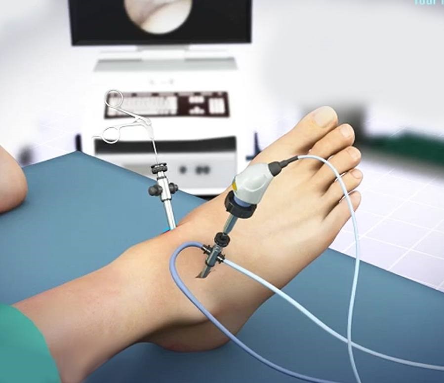
Ankle arthroscopy is a key-hole surgical procedure used to diagnose and treat problems inside the ankle joint. It involves inserting a thin fiber-optic camera (arthroscope) through tiny incisions. The arthroscope transmits high-definition live images to a video screen, allowing the surgeon to visualize internal structures clearly and repair damaged tissues using specialized miniature instruments.
Conditions Treated with Ankle Arthroscopy:
- Arthroscopic Ankle Fusion: For end-stage arthritis cases.
- Arthroscopic Cheilectomy: For anterior or posterior ankle impingement bone spurs.
- Arthroscopic ATFL Reconstruction: Repair of torn lateral ankle ligaments for chronic instability.
- Arthroscopy-Assisted Fracture Fixation: Precise joint surface realignment after trauma.
- Syndesmosis Debridement & Stabilization: Treating high ankle sprains and ligament tears.
“Because arthroscopy uses keyhole micro-incisions, healing occurs much faster, the risk of infection drops significantly, and patient rehabilitation begins earlier.”
What is Subtalar Arthroscopy or Hindfoot Arthroscopy?
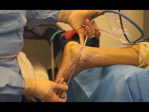
Subtalar arthroscopy is a minimally invasive keyhole technique used to visualize the joint immediately below the ankle joint (the subtalar joint). It is commonly utilized to perform:
- Subtalar joint fusion in severe arthritis cases.
- Arthroscopy-assisted calcaneal (heel bone) fracture fixations.
- FHL (flexor hallucis longus) tendon or Os-trigonum syndrome debridement.
What is Tendoscopy?
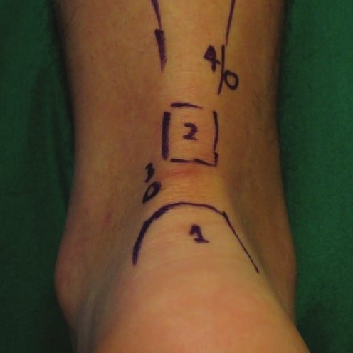
Tendoscopy involves endoscopic visualization inside the tendon sheath using a miniature camera to treat tendon conditions with minimal surgical trauma:
- Achilles Tendoscopy: Performed to excise Haglund’s deformity bumps and treat chronic retrocalcaneal bursitis or Achilles tendinitis.
- Peroneal Tendoscopy: Used to manage outer ankle peroneal tendon inflammation, tears, and subluxation/dislocation issues.